Lung Surgery: Your Evaluation and Tests
You are having lung surgery. Tests and scans may be needed to evaluate your lungs and nearby areas before surgery. You may have already had some of these tests. Others may be scheduled before your surgery. Your healthcare provider uses the information gathered during these tests to help plan your surgery and treatment.
Imaging tests
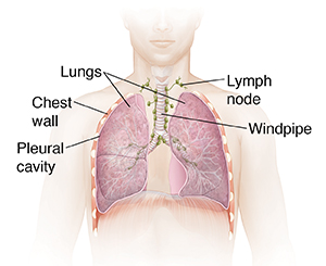
Imaging tests take pictures of your lungs. They allow your healthcare provider to see the inside of your body. They can show problems, such as a mass or tumor, infection, or air in the space between the lungs and the chest wall (pleural cavity). But they can’t tell the healthcare provider for certain whether a change in the lung is cancer. Imaging tests you may have include:
-
Chest X-rays
-
CT scans
-
MRI
-
PET scans may be combined with a CT scan. This is called a PET/CT scan.
-
Other imaging tests as needed, like a bone scan
Tests that look at your lungs (or nearby areas) or take a tissue sample
You may need a test that allows the healthcare provider to look at the inside of your lungs and the area around them. Or you may need to have small samples of lung cells or tissue taken out for testing (called a biopsy). Possible tests used to do this include:
-
Bronchoscopy. This is done using a thin, lighted tube called a bronchoscope. It's put in through your nose or mouth and advanced down into your lungs. It's used to look at breathing passages at the entrance to your lungs. There's a tool within the bronchoscope that can be used to do a biopsy.
-
Mediastinoscopy. A thin, lighted tube (mediastinoscope) is put through a small cut (incision) above the sternum (breastbone). It's used to look at the space between the lungs. A biopsy can be done through this tube.
-
Mediastinotomy. The lymph nodes and other tissues in the chest are looked at by putting a flexible, lighted tube (mediastinoscope) in through an incision in the chest wall next to the breastbone between two ribs. A biopsy may be done. The main difference between this procedure and a mediastinoscopy is the size of the incision and where the tube is placed.
-
Needle aspiration or biopsy. A needle is put through the chest wall into the changed lung tissue. It's used to collect tissue or fluid for testing.
-
Endobronchial ultrasound. A bronchoscope is put down into the windpipe, and a tiny ultrasound device on the end is used to send images of the structures between the lungs to a computer screen. A needle can also be passed down the bronchoscope to do a biopsy if anything abnormal is seen.
-
Thoracentesis. Fluid is removed from the space between the lung and the wall of the chest. It is checked for cancer cells in a lab.
Other tests
You may have tests to measure how well your lungs work or to check for cancer cells. They include:
-
Pulmonary function tests. These measure how well your lungs work. This includes testing how much air your lungs can hold and how much air is left in your lungs after you breathe out (exhale). They can also measure how fast you can blow air out of your lungs.
-
Sputum cytology. You collect sputum a few days in a row. It's sent to a lab and checked for cancer cells.
-
Arterial blood samples. A thin needle can be used to take blood out of an artery in your wrist. This is done to see how much oxygen is in your blood.
© 2000-2024 The StayWell Company, LLC. All rights reserved. This information is not intended as a substitute for professional medical care. Always follow your healthcare professional's instructions.